INSIDE THE BRAIN – Part 3: When the Brain Reveals Its Function Through Failure
- Marcela Emilia Silva do Valle Pereira Ma Emilia
- há 5 dias
- 22 min de leitura


Failure as a Mirror
In the first two parts of this series, we explored the brain from within — its anatomy, its lobes, its integrated systems, its evolutionary history, and its most fundamental cellular unit: the neuron.
But there is a particular way in which the brain has been understood that science has discovered almost by accident. And it was not by looking at the healthy brain that neuroscience advanced most rapidly.
It was by looking at the brain that had failed.
This was meant to be a post about how the entire system presented so far maintains itself in equilibrium and preserves the internal balance of the organism. However, before that, there is something that needs to be explained in order for a function to become visible.
Every lesion, syndrome, and neurodegenerative disease functioned — and still functions — as an unexpected window into discovering a function that existed before the failure. It is almost paradoxical: we only fully understand what a region does when it stops working. That is to say:
🧠 The brain only fully reveals what it does when something stops functioning.
And it is from this logic — from careful clinical observation, from impossible cases, from stories that defied any previous explanation — that much of what we now call modern neuroscience was born.
Here, then, I will present a different part of our series. We are not going to explore what the brain does when it is well. We are going to explore what it reveals when it fails.
🧠 What Are Brain Lesions?
First and foremost, it is important to understand what is meant by a brain lesion and why it is so informative.
A brain lesion is any structural damage to nervous tissue or alteration in its functioning (in any part of the brain or central nervous system — CNS) — whether caused by a stroke (when blood flow is interrupted and neurons die from lack of oxygen), a traumatic brain injury (a direct physical impact), a tumor that compresses or destroys tissue, an infection that inflames the nervous system, or by degenerative processes that progressively destroy specific groups of neurons.
What makes lesions so "valuable" from a scientific standpoint is the principle of functional localization: the discovery that different regions of the brain process different functions. When a specific area is damaged, the functions associated with it are compromised — and so the pattern of deficit, the loss of stable function, reveals with surgical precision what that area was doing.
✨ It is like discovering what a part of an engine does by removing it and observing what stops working.
This logic — which connects directly to the lobes and systems explored in Part 1 of this series — was the foundation upon which neuroscience built its functional map of the brain.
🧠 Function Revealed in Failure
"Nature has sought to ensure that the brain remains healthy. In structural terms, the skull provides thick, protective coverage. The distribution of arteries is extensive and, in much of the brain, even redundant. Even so, the brain is subject to numerous diseases, and the immediate treatment of these conditions is often essential to avoid chronic and debilitating problems or death." (Gazzaniga, Michael S., Ivry, Richard B., Mangun, George R., 2002)
Frontal Lobe — The Case of Phineas Gage

It was 1848, in Vermont, United States, when Phineas Gage, a 25-year-old railway worker, described by his colleagues as responsible, balanced, competent and of good character. A man that people liked to have around. In a workplace accident involving explosives, an iron rod nearly one meter in length pierced his face and skull — entering through the left cheek and passing straight through the left frontal lobe, exiting through the top of his head.
Incredibly, Phineas survived this accident, and it was precisely his survival that changed the history of neuroscience.
Physically, Gage recovered as though nothing had happened. He walked, spoke, saw, moved perfectly — his memory was intact and his intelligence, apparently preserved.
But the Phineas who survived were not the same Phineas as before.
The man previously described as responsible and balanced became impulsive, irresponsible, incapable of planning for the future, and indifferent to the social consequences of his actions. His emotions became unstable. His character — that which people recognized as a unique personality — had disappeared.

🧠 The physician in the case of Phineas Gage, Dr John Martyn Harlow, was the primary doctor to treat Phineas and report these behavioural differences. However, it was only in 1994, with António Damásio and his wife Hanna Damásio (neuroscience researchers), that they were able to provide the first clinical demonstration that the frontal lobe, and in particular the prefrontal cortex, is essential for what is called personality, decision-making, social behaviour, inhibitory control, and planning.
Before Gage, it was believed that personality was something immaterial, disconnected from the brain. After Gage, it became clear: personality has a neurological address.
And it was from this deeper lesson of his case that we have the most widely accepted theory of the "self" — our identity, our character, our way of being in the world.
The theory of the "Self" by António Damásio, which emerged from this specific case, proposes that consciousness and identity arise from the biological regulation of the body, thereby rejecting the theory that separates body from mind. The "Self" is a continuous process, not a fixed entity, constructed through background feelings and emotions, wherein the brain constantly maps the state of the body to create the subjectivity of who the individual is.
The Phineas Gage case shows that specific neurons, in specific regions, when damaged, can make us unrecognizable — even to ourselves.
Parietal Lobe — Hemispatial Neglect and the World That Disappears
Imagine waking up one day and half the world simply ceasing to exist.
It is not blindness — the eyes are functioning — and it is not paralysis, as the limbs respond. But the left side of space, of the body, of the plate of food in front of you, of the page you are reading... simply disappears from your attention.
This is hemispatial neglect — one of the most fascinating and intriguing syndromes in neurology, resulting most frequently from lesions in the right parietal lobe, especially following a stroke.

Patients with hemispatial neglect do not consciously ignore the left side — they genuinely do not "see", in a sense, what is there. When asked to draw a clock, they place all the numbers only on the right side or on the left side. When they eat, they leave half the plate untouched without noticing. When getting dressed, they do not dress on the left side of the body, dressing only the right side using only the right side of the body. When describing an imaginary square, they only describe what is to the right or left, depending on where the lesion occurred.
Remarkably, this lesion shows that one side is ignored not due to a lack of vision but due to a lack of attention. Indeed, the patient may also present with hemiplegia, paralysis of the left hand due to physical paralysis.
🧠 What hemispatial neglect reveals is that spatial attention is not a passive process — having eyes is not enough to see. The brain must actively direct attention towards space, and this capacity depends critically on the parietal lobe, in particular the non-dominant hemisphere.
✨ Seeing is not merely a matter of eyes. It is a matter of the brain.
Hemispatial neglect is therefore not a purely visual problem, but rather the brain ignoring information coming from that side of the brain.
However, there is still treatment for this condition. This condition is commonly the result of a stroke — cerebrovascular accident — and its treatment aims to reduce the failure in environmental perception by focusing on restoring attention and compensating for limitations. Mirror Therapy, Brain Stimulation (tDCS), Prismatic Adaptation, and visual scanning training are the most effective approaches.
Treatment does not offer a rapid or definitive cure, but rather a significant improvement and rehabilitation of the lost capacity. Cerebral neuroplasticity is the key to compensating for the damaged area and restoring the greatest possible functionality and autonomy to the patient.
Temporal Lobe — The Language That Is Lost and the Memory That Does Not Record
The temporal lobe holds two of the most striking cases in the history of neurology — one about language, another about memory.
Wernicke's Syndrome
At the end of the 19th century, German neurologist Carl Wernicke described a pattern of lesion that produced something deeply disturbing: patients who spoke fluently, with normal intonation, with grammatically perfect and structured sentences, but whose words and sequence made no sense.
It was like listening to a speech in which all the words are understood, but the sentences have no meaning whatsoever: "The blue sings the table of the wind when the chair sleeps."
Lesions in Wernicke's area — a region located in the left temporal lobe — impair language comprehension, and the patient begins to speak nonsense whilst also failing

to understand what others say. And, crucially, they often do not realize that what they say makes no sense.
🧠 In contrast, Broca's syndrome — a lesion in the left frontal lobe, crucial for speech production, word articulation, and language processing — revealed that language is not a function of a single brain region, but of a distributed system with specialized components in distinct regions.
✨ Speaking and understanding are cerebrally separate processes.
Aphasia, the condition affecting these areas and which was first brought to wider attention through the case of Bruce Willis, involves impairment of either Wernicke's area, affecting language comprehension and the meaning of what is spoken; or Broca's area, causing difficulty in speech production (slow, effortful, non-fluent speech) whilst comprehension remains preserved; or of both areas, drastically impairing the production, comprehension, and ability to communicate clearly.
The Case of H.M. — The Man Without Memory

In 1953, a young American named Henry Molaison — widely known only by his initials H.M. — underwent an experimental surgery to treat severe epilepsy.
The neurosurgeon bilaterally removed a large part of the hippocampus and adjacent structures of the medial temporal lobe.
The result? The epilepsy improved. But the price was devastating.
When H.M. awoke, he had lost the ability to form new long-term memories. He could converse, reason, laugh, solve problems — but minutes after the conversation ended, there was no trace of what had happened. Each time researcher Suzanne Corkin — for approximately 50 years — entered the room, it was the first time they had ever met. Every single day.
H.M. lived this way for 55 years, until his death in 2008.

🧠 What the case of H.M. revealed is that the hippocampus is essential for the consolidation of new memories, primarily declarative ones — the transfer of information from short-term to long-term memory. That is to say, without this small and specific piece of the brain, the present never becomes the past.
But something even more fascinating was revealed: although H.M. could not create long-term memories, he was able to learn new motor skills. It was like teaching him to ride a bicycle one day — but the following day he had no recollection of having learnt the task the day before, yet when he went to perform it, his brain had retained the motor information.
✨ It was then discovered that there were two separate memory systems within the same brain: one for facts and events, another for skills and procedures.
This case, together with that of Phineas Gage, is considered by many to be among the most astonishing in the history of modern neuroscience.
Occipital Lobe — Eyes That See, but a Brain That Does Not Process
The occipital lobe is entirely dedicated to visual processing, and lesions in this area produce some of the most disturbing and counterintuitive experiences in neurology.
Cortical blindness occurs when the primary visual cortex is damaged — the eyes are perfectly functional, the optic nerve intact, yet the patient cannot see. Visual information reaches the brain, but is not processed.

Even more remarkable is the phenomenon of blindsight: patients with cortical blindness who are able, when tested, to correctly point to objects they are asked to locate, yet insist they cannot see them. That is to say, the brain processes visual information unconsciously, without any conscious perception.
What these occipital lesions reveal is that seeing is a process of active construction involving various parts of the occipital lobe. The brain does not passively receive images — it constructs, interprets, and organises them. When this construction fails at any stage, visual reality disappears, even with perfectly functioning eyes. The person may lose their vision entirely, or "see without seeing".
The case of visual agnosia — the inability to recognise objects, people, or colours visually, despite being able to see them — demonstrates in practice that recognition and vision are separate processes. A patient with agnosia may see an object but be unable to identify what it is, and in more severe cases may not even be able to copy it in a drawing if asked to do so.
Treatment consists of rehabilitation with occupational therapists to help awaken and sharpen the other senses, such as touch and hearing, to compensate for the visual loss. Unfortunately, there is still no direct cure.
✨ Seeing is not enough. One must recognize. And recognizing is a cerebral function, not an ocular one.
Limbic System — Klüver-Bucy Syndrome and the End of Fear
In 1937, researchers Heinrich Klüver and Paul Bucy bilaterally removed the amygdala and other structures of the medial temporal lobe in rhesus macaques.
The result was bewildering.
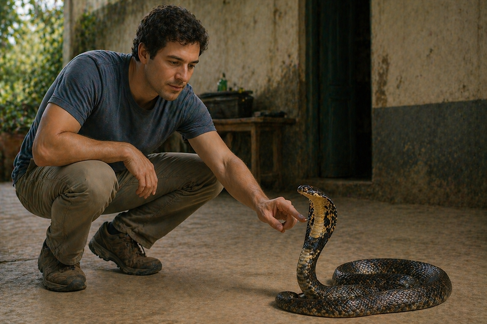
Animals that had previously reacted with aggression and fear to threatening stimuli — such as a snake or an unfamiliar human — became entirely unperturbed. Without fear. Without aggression. Without the capacity to assess danger.
But there was more: the animals developed hyperorality (placing everything in their mouths), hypersexuality, an inability to recognize the emotional significance of objects, and a strange tendency to visually examine everything with great intensity, yet without being able to interpret what they saw.
🧠 Klüver-Bucy syndrome revealed that the amygdala is central to the processing of fear, threat, and the emotional significance of stimuli. Without it, the world loses its affective valence — nothing is dangerous, nothing is threatening, nothing activates the alerting response that keeps animals alive.
However, the syndrome can arise unexpectedly, without the removal of any part of the brain, emerging instead from damage to both temporal lobes, or it may be caused by neurodegenerative diseases (such as Pick's Disease and Alzheimer's), herpetic encephalitis, cranial trauma, tumors, or cerebrovascular accidents. Even so, it is important to acknowledge that the condition is rare among humans, being more documented in studies with primates.
There is no cure for this condition, and its treatment aims to alleviate the symptomatic behaviours.
✨ Fear is not weakness. It is an essential cerebral function for survival.
🧬 Neurodegenerative Diseases — When the Brain Loses Itself
If focal lesions — whether of external or unexpected origin — reveal the function of specific regions, neurodegenerative diseases reveal something even more shocking: what happens when the brain begins to destroy itself in a progressive and irreversible manner.
These are diseases that still have no cure, that advance slowly, and that, as they progress, reveal — layer by layer, leaving a trail of destruction — what each brain system was sustaining.
Alzheimer's — The Memory That Dissolves Identity
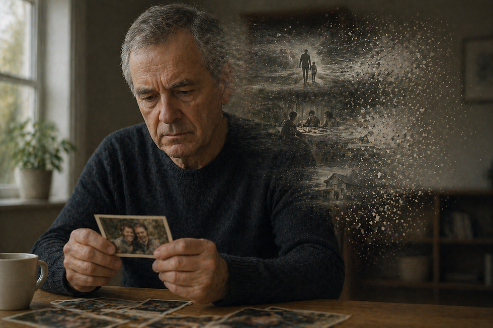
Alzheimer's disease is the most common form of dementia, affecting more than 55 million people worldwide. It begins silently — small lapses in memory, difficulty finding words, occasional disorientation.
But do not be frightened or alarmed by these initial symptoms, for beneath them a biological cascade has already been underway for years: such as the accumulation of beta-amyloid plaques between neurons and of tau protein tangles within them, which are progressively destroying synaptic connections and killing neurons. Symptoms that require a diagnosis.
The hippocampus — that same region from the case of H.M. — is one of the first regions affected, and it is for this reason that difficulty forming new memories is frequently the first sign.
🧬 As the disease progresses, the prefrontal cortex, the temporal cortex, and other associative regions are progressively compromised. With the symptoms advancing, long-term memory fades — first forgetting recent events, then older ones. Language deteriorates — aphasia is one of the complications of Alzheimer's. The recognition of familiar faces disappears — prosopagnosia is one of the most evident signs of the disease. And the individual's personality becomes lost along the way.
✨ Alzheimer's is not merely a disease of memory. It is a disease of identity.
The progression of Alzheimer's is deeply distressing, yet it reveals the hierarchy of memory in the brain — what is stored where, and in what order it is lost. It also reveals how personal identity is not an abstract entity, but depends on specific neural networks which, when destroyed, take with them what was once called the "Self".
In a sense, one might say that, as painful as the disease is, in many cases its progression alters the perception of one's own condition, which may modify the way in which suffering is experienced.
Find out more about what life with Alzheimer's is like in this article:

Parkinson's — The Body That Freezes
Parkinson's disease often begins with almost imperceptible symptoms: a slight tremor in the hand at rest, a stiffness in movement, a slowness that people attribute to ageing.
However, beneath these signs, a progressive destruction of the dopaminergic neurons of the substantia nigra is taking place — a structure in the basal ganglia, those deep brain structures that control voluntary movement, the initiation of action, and motor fluidity.

The progressive loss of neurons in the substantia nigra of the brain, the area responsible for producing dopamine, causes an interruption in neuronal communication. Without this neurotransmitter, associated with motivation, reward, and the modulation of movement, movements become slow (bradykinesia), rigid, and tremulous. In more advanced stages, the patient may freeze mid-step, unable to initiate or complete the movement.
🧠 Parkinson's revealed the central role of the basal ganglia in the fluidity and control of movement — something that had previously been largely attributed solely to the cerebellum and the motor cortex.
✨ Each tremor of a patient with Parkinson's is, in fact, the brain attempting to communicate something that healthy neurons are doing in silence: controlling movement without one having to think about it.
Furthermore, it also revealed the fundamental role of dopamine — not only in movement, but in motivation, pleasure, and reinforcement learning.
Parkinson's, like Alzheimer's, still has no cure. However, its symptoms can now be better managed, with dopamine-based medications, primarily L-dopa, and even brain implants in more severe cases, which significantly improve the patient's quality of life.
Actor Michael J. Fox recounts his battle against the disease in this article:

Multiple Sclerosis — When Myelin Disappears
In Part 2, the importance of the myelin sheath was shown in detail — the biological insulation that coats the axon and enables saltatory conduction, making the transmission of the nerve signal up to 60 times faster.
In multiple sclerosis (MS), an autoimmune disease, the immune system itself attacks and destroys the myelin of the central nervous system — in a process called demyelination.

The result is the transformation of rapid nerve transmission into something slow, interrupted, and inefficient. Symptoms vary according to the regions affected: blurred or double vision, extreme fatigue, motor difficulties, tingling, balance problems, and cognitive difficulties.
🧬 MS is especially cruel because relapses and remissions, which are frequent, create an illusion of recovery, and they affect young people, typically between the ages of 20 and 40.
✨ Multiple sclerosis is, in a sense, the most visceral demonstration of the importance of myelin as described in Part 2. The speed that made the neuronal signal so efficient — up to 120 m/s — collapses when the insulation disappears.
And what MS revealed to science goes beyond myelin: it showed that the immune system can become an enemy of the nervous system itself, opening up an entire field of research into neuroinflammation and neurological autoimmune diseases.
Currently, MS has no cure, but modern treatments with immunomodulators and monoclonal antibodies slow the progression of the disease, reducing relapses by 70–80%, thereby improving the individual's quality of life. It is also important that diagnosis be made as early as possible, as immediate treatment prevents accumulated neurological damage.
Amyotrophic Lateral Sclerosis (ALS) — The Silence That Advances

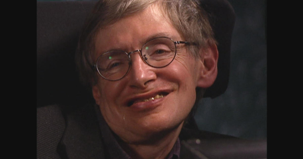
Amyotrophic lateral sclerosis (ALS) — known internationally as the disease of Stephen Hawking — is one of the most devastating diseases in existence. It is to live inside a body that is dying.
It specifically affects motor neurons — those that send signals from the brain and spinal cord to the muscles, those responsible for voluntary and involuntary movements. As these neurons die, the muscles are progressively deprived of stimulation, atrophying and becoming paralyzed.
Progressively, the innate capacity to walk, to swallow, to speak, to breathe is lost. The mind, in the majority of cases, remains intact — which makes ALS particularly cruel: the person is present, conscious, yet imprisoned within a body that is gradually ceasing to respond.
🧠 What ALS revealed was the anatomical separation between the motor and cognitive systems — and the absolute dependence of the body on motor neurons for any expression of life in the physical world.
Amyotrophic lateral sclerosis is unfortunately a disease without cure, neurodegenerative and fatal. Its treatment focuses on slowing progression by controlling symptoms and adapting the environment to improve the patient's quality of life. The prognosis is challenging, and generally unfavorable due to the progressive nature and rapid degeneration of motor neurons, which leads to paralysis and respiratory difficulties within 3 to 5 years of diagnosis.
To learn more about the disease, Stephen Hawking's story was depicted in the film "The Theory of Everything", available on Netflix and Prime Video, as well as a documentary featuring him on Apple TV, "Genius by Stephen Hawking", and this exceptional interview with him:
🧩 Rare Syndromes — The Cases That Challenged Science
Beyond lesions and diseases, neurology is also the territory of rare, extraordinary syndromes that seem drawn from a novel — and which revealed, precisely through their strangeness, profound aspects of how the brain constructs reality.
Rare diseases and syndromes represent one of the greatest challenges in the neurological field of study, frequently leaving scientists and physicians without clear answers regarding causes, progression, or cure. Many of these conditions are now known to be genetic, degenerative, or autoimmune, evolving in peculiar ways that defy current knowledge.
Capgras Syndrome — The Impostors Who Took the Place of Loved Ones

Imagine looking at your mother and being absolutely certain that she has been replaced by an impostor. The face is identical, the voice is the same, the gestures are familiar — but it is not her. It is a copy. A substitute.
This is Capgras syndrome, first described in 1923 by French psychiatrist Joseph Capgras.
Visual recognition remains intact — the patient knows that that face belongs to their mother. But the emotional response of familiarity is absent; the brain, confronted with this mismatch between recognition and emotion, constructs the only explanation that seems to make sense: that cannot be their mother. It must be a copy.
🧠 The delusion is not a failure of reasoning. It is a desperate attempt to explain a genuinely strange neurological experience.
In this case, the patient is not fabricating an excuse for what they are feeling, but is trying to explain the reality of what they are truly experiencing. The strangeness regarding the person before them does not lie in general recognition, but is associated with the absence of the emotional bond that the patient does not feel towards that individual.
What neuroscience has discovered is fascinating: the recognition of faces and the emotional response to those faces are two separate processes. Normally, upon seeing the face of a loved one, the brain not only recognizes them visually — it also generates an automatic emotional response of familiarity and affection.
✨ Capgras syndrome revealed that the identity of the people we love is not merely visual — it is emotional. And that when the emotional circuit fails, the brain would rather "invent" impostors than accept that something within itself is not working.
Cotard Syndrome — The Conviction of Being Dead
If Capgras syndrome is disturbing, Cotard syndrome is even more bewildering.

Described in 1882 by French neurologist Jules Cotard, this syndrome is characterized by the delusion in which the patient themselves believes they are dead, that they do not exist, that they have no organs, no blood, that they are rotting away.
In an extreme manifestation of the disease, patients with Cotard syndrome refuse to eat — because the dead do not eat. They assert that their heart does not beat, that their blood does not flow, that they no longer exist.
Modern neuroscience does not yet have a complete explanation for this syndrome, but associates Cotard syndrome with dysfunctions in the prefrontal cortex and the limbic system, particularly the amygdala. It is frequently associated with severe depression or schizophrenia due to its nihilistic delusions, apathy, and isolation. Its treatment therefore involves a combination of antidepressants, antipsychotics, and psychotherapy.
The hypothesized cause of the syndrome is similar to that of Capgras: there is a dissociation between the perception of one's own body and the emotional response of familiarity, now, with one's own body.
This disease is one of the most extreme examples of how the brain is, fundamentally, a constructor of reality — and of how that reality can collapse in ways impossible to imagine when the right circuits fail.
Alien Hand Syndrome — The Arm That Does Not Obey
Alien hand syndrome is exactly what the name suggests: the patient feels that one of their limbs — typically the hand — does not belong to them, acts autonomously, and often works against their own will.

The "alien" hand may pick up objects without the patient wanting it to, unbutton buttons that the other hand has just buttoned, or even attempt to strangle the patient themselves. These are truly involuntary, complex, and autonomous movements, acting as though it had a will of its own.
🧠 What this syndrome reveals is that the two hemispheres have relatively independent motor control systems, and that the sensation that movements are intentional and our own depends on a continuous integration between these systems.
✨ The experience of "wanting" to move one's arm — which seems the most natural and obvious thing in the world — is, in fact, the result of a complex cerebral coordination which, when it fails, produces limbs that appear to have a will of their own.
It occurs typically following lesions to the corpus callosum — the great communication pathway between the two cerebral hemispheres — or to the medial frontal lobe. But it may also result from other brain lesions such as cerebrovascular accidents (CVA), tumors, and neurodegenerative diseases.
Prosopagnosia — A World Without Faces

Prosopagnosia — or face blindness — is the inability to recognize familiar faces, even the closest ones. Patients with severe prosopagnosia may be unable to recognize their spouse, their children, or their own face in the mirror.
Vision is intact, faces are perceived, but the specific recognition of facial identity — that which makes your mother's face unmistakable — is absent. The person cannot identify changes in people's appearance or even interpret facial expressions.
🧠 Prosopagnosia revealed the existence of a specific brain region for face recognition: the fusiform face area (FFA), located in the fusiform gyrus of the inferior temporal lobe.
✨ Faces are so important to human social survival that the brain evolved a specific region solely to process them.
Prosopagnosia showed that face recognition is not a generic visual function — it is a specialized system, separate from object vision, that can be selectively destroyed by highly specific lesions.
The causes of this condition are not yet fully understood, but it is known that it can be congenital (genetic/developmental) or acquired through brain lesions such as stroke, tumors, or trauma to the right hemisphere.
Diagnosis is made through neuropsychological tests and, unfortunately, there is no known cure for this condition. However, treatment involves developing other compensatory sensitivities, such as recognizing others by their voice, hair, clothing, or manner of walking.
Great care is also urged with individuals who have this condition, as their mental state requires attention to the extent that these individuals may experience social and professional difficulties, often appearing distant or disinterested. This is not their fault, but the fault of the disease itself, which can also trigger anxiety crises and depression, if those around them are not well informed.
Oliver Sacks, a renowned neurologist, became famous for humanizing clinical cases of prosopagnosia, thus becoming a celebrated British writer. He himself suffered from the condition, and his books in a sense reflected his own reality. Suggested reading: his book "The Man Who Mistook His Wife for a Hat", but there is also this interview in which he speaks about his difficulties: https://www.npr.org/transcripts/130732146, or alternatively the documentary "Oliver Sacks: His Own Life", available on Apple TV or Prime Video.

🌱 Neuroplasticity — The Brain That Reorganizes Itself
The brain is a marvelous machine, which survives even under adverse conditions. Lesions, syndromes, diseases — it would be easy to paint a picture of a fragile, deterministic brain, devoid of any capacity for recovery.
But the brain is marvelous! The brain has learnt to adapt; in fact, the brain is plastic.
Neuroplasticity is the capacity of the nervous system — more specifically its neurons — to modify their structure and function in response to experience, learning, environment — and, of course, damage.
Following a brain lesion, the brain does not simply accept the loss: it reorganizes itself so that adjacent regions may assume, partially or permanently, the functions that belong to damaged areas. New synaptic connections are formed, broadening the function of specific neural circuits and neurons that have already been consolidated.

There are cases of remarkable recovery following stroke that demonstrate this in impressive fashion: patients who completely lost speech and recovered language after years of intensive rehabilitation. Children who, following hemispherectomies (removal of an entire hemisphere), developed language and mobility in the remaining hemisphere.
Yes, neuroplasticity is greater in childhood — the young brain is extraordinarily adaptable — but this does not mean it disappears in adulthood. Throughout life, the brain continues to modify itself in response to what we experience and learn.
✨ Neurological rehabilitation is, at its core, a conversation with the plasticity of the brain — an invitation for it to reorganize what can be reorganized.
And perhaps it is here that the story of lesions and diseases becomes even more surprising: it is not only the failure, but the extraordinary capacity of some recoveries — partial, slow, difficult — yet the brain nonetheless manages to adapt in order to survive.
To learn more about neuroplasticity, visit the post

🌍 Conclusion — Vulnerability as Wisdom

The history of discovery and confrontation with neurological diseases spans almost two centuries — not all through planned laboratory experiments, but through careful observation of people who suffered, who lost functions, who became clinical cases without having chosen to.
Phineas Gage did not choose to be the case that revealed the frontal lobe. H.M. did not choose to be the case that mapped the hippocampus and memory. The patients with Alzheimer's, with Parkinson's, with ALS, with the rare syndromes we explored — none of them chose to be a window into neuroscience.
And it is for this reason that these stories deserve to be told with care, with respect, and with the awareness that behind every clinical case lies an entire life.
🧠 What we learn from the failure of the brain is not merely science. It is also a profound lesson about what we are.
We are memory — and when it disappears, identity goes with it. We are emotion — and when circuits fail, reality transforms into something unrecognizable. We are movement, language, attention, recognition — and each of these capacities depend on specific neural networks, which can be destroyed by a blood clot, by an iron rod, by a misfolded protein.
✨ Knowing the vulnerability of the brain is not cause for despondency. It is, paradoxically, an invitation to care — to sleep, to movement, to relationships, to cognitive stimulation, to the management of stress.
And it is also an invitation to humility: the "self" that each of us believes ourselves to be — stable, continuous, recognizable — is, in fact, a dynamic construction of neural networks working in silence, every day, to keep us coherent with ourselves.
🧠 In the next part, I will leave the realm of failure and truly enter the theme of balance, exploring how the brain maintains life through homeostasis, and the sympathetic and parasympathetic systems. Because after understanding how the brain can fail, it is time to understand how, for most of the time, it keeps us alive without us even noticing.
📚 References and Essential Reading
Damasio, A. Descartes' Error: Emotion, Reason and the Human Brain. Vintage, 2006.
Sacks, O. The Man Who Mistook His Wife for a Hat. Picador, 1985.
Ramachandran, V. S. Phantoms in the Brain: Probing the Mysteries of the Human Mind. William Morrow, 1998.
Corkin, S. Permanent Present Tense: The Unforgettable Life of the Amnesic Patient H.M. Basic Books, 2013.
Nolte, J. The Human Brain: An Introduction to Its Functional Anatomy. Elsevier, 2008.
Cosenza, R. M. Fundamentos de Neuroanatomia. 4th ed. Guanabara Koogan, 2021.
Macmillan, M. (2000). An Odd Kind of Fame: Stories of Phineas Gage. MIT Press.
Baddeley, A., Aggleton, J. P., & Conway, M. A. (Eds.). (2002). Episodic Memory: New Directions in Research. Oxford University Press.
Klüver, H., & Bucy, P. C. (1937). "Psychic blindness" and other symptoms following bilateral temporal lobectomy in rhesus monkeys. American Journal of Physiology, 119, 352–353.
Cotard, J. (1882). Du délire des négations. Archives de Neurologie, 4, 152–170.
Wilgus, J., & Wilgus, B. (2009). Face to face with Phineas Gage. Journal of the History of the Neurosciences, 18(3), 340–345. https://doi.org/10.1080/09647040903018402
Van Horn, J. D., et al. (2012). Mapping connectivity damage in the case of Phineas Gage. PLOS ONE, 7(5), e37454. https://doi.org/10.1371/journal.pone.0037454
Augustinack, J. C., et al. (2014). H.M.'s contributions to neuroscience: A review and autopsy studies. Hippocampus, 24(11), 1267–1286. https://pmc.ncbi.nlm.nih.gov/articles/PMC6007845/
👉 Further Reading
Phineas Gage's Great Legacy: https://pmc.ncbi.nlm.nih.gov/articles/PMC7735047/
The Legacy of Patient H.M. for Neuroscience: https://pmc.ncbi.nlm.nih.gov/articles/PMC2649674/
Alzheimer's Disease International: https://www.alzint.org/about/dementia-facts-figures/dementia-statistics/
The Return of Phineas Gage: Clues About the Brain from the Skull of a Famous Patient (António and Hanna Damásio study): https://ubneuro-ccohan.webapps.buffalo.edu/readings/PhineasGage.pdf
NHS Parkinson's Information Website: https://www.nhs.uk/conditions/parkinsons-disease/
Associação Brasileira de Alzheimer: https://abraz.org.br/


Comentários